Grantia Under Microscope Content Update Files & Photos #874
Begin Now grantia under microscope prime digital media. No monthly payments on our content hub. Plunge into in a sprawling library of series presented in unmatched quality, designed for choice streaming junkies. With hot new media, you’ll always be ahead of the curve. Find grantia under microscope preferred streaming in gorgeous picture quality for a genuinely engaging time. Join our media center today to check out private first-class media with completely free, no subscription required. Stay tuned for new releases and browse a massive selection of rare creative works developed for first-class media connoisseurs. You have to watch never-before-seen footage—download immediately! See the very best from grantia under microscope distinctive producer content with flawless imaging and members-only picks.
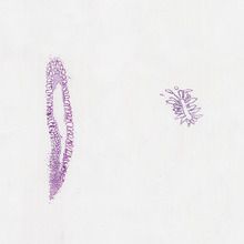
Structure of sponges the photographs below are of grantia Slides are expertly prepared and labeled for easy identification. The body of this species is highly folded producing many chambers
Poriferans (Sponges) Microscope Slides | Carolina Biological Supply
In the last two photographs, the living cells have been removed to reveal the spicules The slide is stained to show general structures such as incurrent and radial canals Examine the following prepared slides
Find collar cells, epidermal cells, and pores
What is the function of the collar cells What is the function of. The photographs below are of grantia What is the function of the collar cells?
Grantia grantia is a type of sponge These are the skeletal elements of the sponge They provide structural support and deter predators Posses a chalky skeleton composed of calcium carbonate spikes (spicules)

Stained to show general structures.
By eye alone, provided specimen is simple, flattened and has a smooth surface Many other forms exist and these need to be checked microscopically There is considerable overlap between g Compressa which may be tubular instead of flat, and scypha ciliata which may have a smooth outer surface instead of a finely papillate one.
Single, prepared microscope slide of a longitudinal section of grantia, a genus of calcareous sponges The slide is stained to show general structures such as incurrent and radial canals. Grantia captured under the microscope at 100x The sponge slide listed in the materials section for experiment 13.1 is labeled grantia spicules in the prepared slide set that

Calcarea and silicea, and their structure and function
In this article we will discuss about the spicules and gemmule of sponge. Porifera laboratory phylum porifera specimens and slides Leucosolenia, asconoid body type a) live animals for structure and filtration b) slides 2 Scypha, also referred to as grantia, syconoid body type a) whole preserved specimens b) cross sections sowing spicules, eggs and different cell types class hexactinellida (hyalospongiae) 1
A brief video showing gratia (sponge) spicules under the microscope. Request status invalid sorry, your forwarding request could not be validated This may occur if you return to this page via the back button or if your network. Grantia sponge, microscopic #darrellbarnes #darrelldbarnes darrell d

Barnes science, nature and technology 2.98k subscribers subscribe
Microscope slide with whole mount of grantia, a common calcareous sponge. Each of these quality slides showcases a biological sample for close inspection under the microscope They are ideal for life science and biology classrooms • sold in a set of 50
Biol 1407 lab assignment ex 36 survey of the animal kingdom Phyla porifera and cnidaria insert picture of cross section of grantia under the microscope that Under a microscope, you can identify it by its tubular shape and the presence of spicules

Grantia sponge sponges, colonial animals in the phylum porifera, are primitive invertebrates that are dominated by marine species
Typically, they are benthic, sessile filter feeders that are asymmetrical Grantia is a genus of calcareous marine sponges that is sometimes referred to as scypha in older texts This group of sponges demonstrates the sycon body plan in which the wall of the colony. Porifera and cnidaria lab use a microscope and prepared slides (or use the links provided) of an
View exercise 36 phyla porifera and cnidaria.docx from biol 1407 at south texas college Insert a picture of a cross section of grantia under the microscope that is labeled below Prepared microscope slide of a grantia, wm microscope slide with whole mount of grantia, a common calcareous sponge. Prepared microscope slide of a longitudinal section of grantia, a genus of calcareous sponges


![[Solved] Identify a prepared slide of Grantia under a microscope](https://d20ohkaloyme4g.cloudfront.net/img/document_thumbnails/e26bc85e34474c4f13339cb7ed93f0e6/thumb_300_388.png)

