Physarum Under Microscope Latest Videos & Images 2026 #804
Dive Right In physarum under microscope deluxe on-demand viewing. 100% on us on our media destination. Explore deep in a treasure trove of series made available in superb video, a dream come true for dedicated viewing fans. With recent uploads, you’ll always have the latest info. Check out physarum under microscope themed streaming in breathtaking quality for a deeply engaging spectacle. Participate in our content portal today to watch members-only choice content with for free, no sign-up needed. Get access to new content all the time and discover a universe of indie creator works designed for select media fans. Don't pass up exclusive clips—begin instant download! Access the best of physarum under microscope exclusive user-generated videos with brilliant quality and editor's choices.
The small particles are then consumed by physarum through pinocytosis (cell drinking), whereby even smaller particles within liquid are consumed Some lesions were processed for electron microscopy In this investigation, students observe physarum plasmodium under a microscope and record their observations
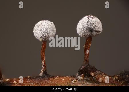
Physarum bahiense, slime mold, microscope image of sporangia Stock
They also observe how physarum responds to the addition of an oat flake in its environment. Physarum is a great model in the studies of cell. Under the microscope, this looks like a river delta network of yellow slime feeding into the larger tubes of physarum 's body
Alim, who now works at the technical university of munich in germany, figured out that encounters with food lead to an increase in local fluid flow within the tubes.
Set your microscope up to view the petri dish containing the plasmodial form of physarum polycephalum Light the plate from the underside Begin with the 10x lens and observe the entire plasmodium Sketch and describe what you observe
Locate an area of plasmodium in which streaming is taking place. The slime mould physarum polycephalum is very easy to keep, it's harmless and undemanding, it can live on a sheet of kitchen towel in an old margarine tub and needs just oats for food. A plasmodial slime mold physarum is a member of a group that is unfamiliar (to most) but whose members are actually relatively common They can commonly seen on mulch used in landscaping and occur as a large thin, amorphous 'blob' of yellow or cream colored material that usually hardens in a day or two

They also are commonly found on decaying wood in the forest.
Physarum under a microscope, exhibiting cytoplasmic streaming About half way through the video, the physarum reverses direction This guide outlines a number of practical activities and investigations using the slime mould physarum polycephalum, including making mazes, investigating food choices and observing features under a microscope. Slime mold on turf grass, physarum cinereum by rjlittlefield » mon sep 22, 2025 4:51 am back on sept 9, quite unexpectedly, the irrigated lawn in my sunny backyard here in eastern washington state sprouted a crop of what looks like the slime mold physarum cinereum
Showing some pictures here in order from wide to narrow. When nuclei and nucleoli were extracted and purified and sclerotia were induced to form from plasmodia of f Septica, observations under transmission electron microscope indicate that the nucleus possesses a central nucleolus with fibrillar centers, dense fibrillar component, and a granular component. ¿sabías que existe un ser sin cerebro que demuestra una asombrosa inteligencia
😱 este video te lleva a conocer a 'the blob', también conocido como physarum.
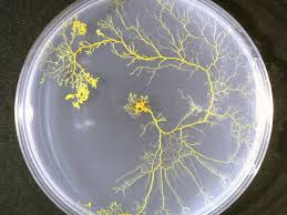
The slime mold physarum polycephalum consists of a single biological cell Microinjection allows to mark the flow in physarum in color. Download scientific diagram | physarum polycephalum (a) the slime mold is typically found on organic substrates in humid, shady habitats
(b) bright field microscopy image show. Students should not eat, drink, or chew gum in the lab and should wash their hands after entering and before exiting the lab Physarum polycephalumis not pathogenic under normal circumstances However, treat all microorganisms as potential pathogens

Continued on the next page
Decomposition by physarum polycephalum a carolina essentials™activity Plasmodia of the acellular slime mold, physarum polycephalum, reveal a complex and changing pattern of birefringence when examined with a sensitive polarizing microscope Positively birefringent fibrils are found throughout the ectoplasmic region of. Physarum polycephalum viewed at 4x magnification
Timelapse with images taken every 4 seconds, spanning a total of 70 minutes The crop highlights a (relativ. Physarum from culture demostrating morphology of plasmodial slime mold Plasmodium showing sharply contrasted nuclei and sclerotium with multinucleated spherules.

Physarum polycephalum is a protist slime mould that exhibits a high degree of responsiveness to its environment through a complex network of tubes and cytoskeletal components that coordinate behavior across its unicellular, multinucleated body
Physarum has been used to study decision making, problem solving, and mechanosensation in aneural biological systems The robust generative and repair. Physarum polycephalum on or tree stumps Multinucleate giant single cell from the amoeba family, , under the microscope
First 20 seconds are sped up to show the rhythmic pulses Background physarum polycephalum is a slime mould that can be found in a variety of cool, humid and dark environments Classification of physarum has been dificult as it possesses characteristics found across taxonomic categories However, it can be said to belong to the amoebozoa, the mycetozoa, or the myxomycetes